House Dust Mite
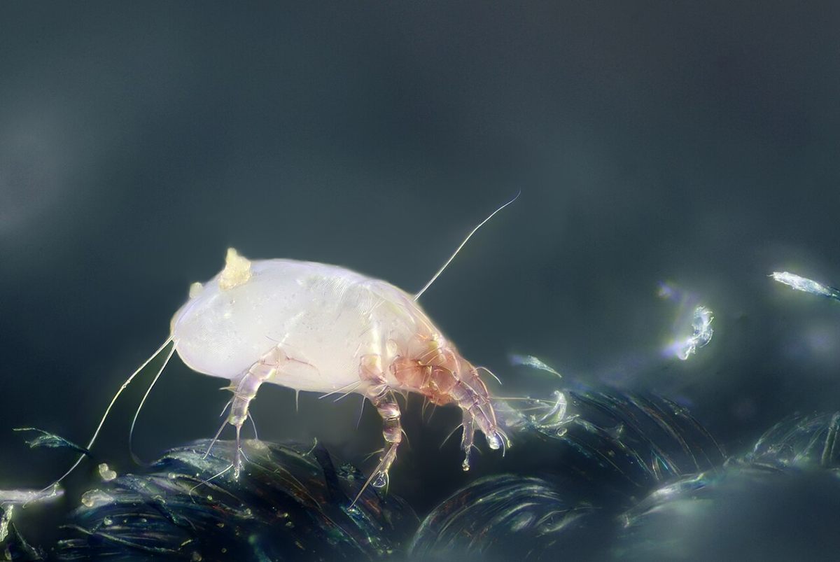
The house dust mite (Dermatophagoides pteronyssinus) is a microscopic arachnid, about 0.4 millimeters long, that lives in bedding, carpets and upholstery and feeds on shed human skin. Its droppings are a major source of indoor allergens.
The house dust mite (Dermatophagoides pteronyssinus) is a microscopic arachnid, about 0.4 millimeters long, that lives in bedding, carpets and upholstery and feeds on shed human skin. Its droppings are a major source of indoor allergens.
🧬 Field Guide Specimen: Open House Dust Mite in the CRV Science Field Guide to log the specimen, see its place among the Ecdysozoa lineages in the Cladogram Explorer, and test your recall in the Red List conservation game.
House Dust Mite at a Glance
| Property | Value |
|---|---|
| Scientific name | Dermatophagoides pteronyssinus |
| Kingdom | Animalia |
| Phylum | Arthropoda |
| Subphylum | Chelicerata |
| Class | Arachnida |
| Order | Sarcoptiformes |
| Field Guide group | Ecdysozoa: Arachnida (Spiders, Scorpions & Allies) |
| Diagnostic feature (Field Guide) | Microscopic translucent oval arachnid with finely striated cuticle and long sensory setae bristles. |
| Size | About 0.4 millimeters |
| Main allergen | Der p 1 (protease) |
Evolutionary Systematics & Comparative Anatomy
Dermatophagoides pteronyssinus is an astigmatid acarine belonging to the family Pyroglyphidae within the order Sarcoptiformes (suborder Oribatida, cohort Astigmata). Phylogenetic reconstructions using nuclear ribosomal genes and complete mitochondrial genomes demonstrate that astigmatid mites—including house dust mites and vertebrate skin parasites such as Sarcoptes scabiei—originated as a derived clade nested inside soil-dwelling oribatid mites. Within Pyroglyphidae, free-living detritivores ancestral to Dermatophagoides inhabited avian nests, feeding on shed feather keratin and dander before transitioning to human dwellings as commensal synanthropes during the rise of permanent agricultural settlements.
Adult D. pteronyssinus are ovoid, cream-colored or translucent arachnids measuring 250 to 320 micrometers in length in males and 350 to 420 micrometers in gravid females. As in other Acari, ancestral arachnid tagmosis is obscured by the fusion of the prosoma and opisthosoma into an unsegmented idiosyncratic body plan divided into an anterior gnathosoma (bearing the trophic appendages) and a larger posterior idiosoma. The gnathosoma carries a pair of two-segmented chelate-dentate chelicerae adapted for scraping and shearing desquamated epidermal corneocytes, flanked by sensory pedipalps.
The cuticle covering the idiosoma is thin, weakly sclerotized, and etched with fine parallel fingerprint-like striations (plicate cuticular ridges) that confer mechanical flexibility while reducing evaporative surface area. On the dorsal propodosoma, D. pteronyssinus bears a narrow sclerotized prodorsal shield, whereas the ventral surface carries sclerotized apodemes (epimera) into which the extrinsic coxal muscles insert. Nymphs and adults possess four pairs of five-segmented walking legs (larvae hatch with three pairs), each terminating in a membranous, stalked pretarsal ambulacral sucker (pulvillus) that enables strong adhesion to textile fibers. Sexual dimorphism is prominent: adult males are smaller, possess a more heavily sclerotized ventral hysterosomal shield, and bear a pair of circular adanal copulatory suckers alongside thickened third or fourth legs used to clasp females.
Physiology, Biomechanics & Sensory Biology
Cutaneous Gas Exchange and Active Water Vapor Absorption
Dermatophagoides pteronyssinus lacks both spiracles and a tracheal system—the morphological state that gives the cohort Astigmata its name—and also lacks a discrete heart or vascular network. All respiratory gas exchange of O2 and CO2 occurs via passive Fickian diffusion across the thin, striated idiosomal cuticle, facilitated by a high surface-area-to-volume ratio and a thin sub-epicuticular lipid layer. However, this permeable cuticular barrier exposes the mite to continuous evaporative water loss into the surrounding air.
Because D. pteronyssinus never drinks liquid water, it maintains osmotic homeostasis through active atmospheric water vapor absorption mediated by paired supracoxal glands situated just above the coxae of the first pair of legs. The supracoxal glands secrete a concentrated, hygroscopic hyperosmotic fluid enriched in K+, Na+, and Cl- ions into an open cuticular gutter (Podapolipus duct/atrial groove) that runs downward toward the prebuccal cavity. When ambient relative humidity exceeds the critical equilibrium activity (CEA)—which ranges from 55% to 73% relative humidity depending on ambient temperature (typically around 60% at 20 °C)—atmospheric H2O vapor condenses into the hypertonic salt solution along the groove. The hydrated fluid is then ingested via the pharyngeal pump, and water is resorbed across the gut epithelium while ions are recycled to the supracoxal glands. Below the CEA, the salt solution crystallizes and clogs the supracoxal openings; the mite reduces metabolic activity, draws its legs inward, and can survive temporary desiccation, though prolonged exposure to <45% relative humidity results in lethal dehydration.
Digestive Enzymology and Molecular Allergenicity
The digestive tract of D. pteronyssinus consists of a muscular pharynx, a narrow esophagus, a ventriculus with paired gastric caeca, a globular colon, and a post-colon where digested material is compacted into peritrophic membrane-wrapped fecal pellets measuring 10 to 40 micrometers in diameter—a particle size comparable to anemophilous pollen grains. Each mite excretes roughly 20 fecal pellets per day, packed with active digestive enzymes and residual protein fragments.
More than 30 distinct allergen groups (designated Der p 1 through Der p 38+) have been characterized from D. pteronyssinus. The major group 1 allergen, Der p 1, is a 25-kilodalton papain-like cysteine protease secreted by the midgut epithelium. When inhaled onto human bronchial mucosa, Der p 1 proteolytically cleaves the extracellular domains of the tight-junction proteins occludin and claudin-1, disrupting epithelial barrier integrity and enabling paracellular penetration of allergens to submucosal dendritic cells. Furthermore, Der p 1 cleaves the low-affinity IgE receptor (CD23) on B lymphocytes and the alpha subunit of the IL-2 receptor (CD25) on T cells, biasing adaptive immunity toward a T helper 2 (TH2) response and triggering IgE-mediated mast cell degranulation (see Immunology and Antibody). A second major allergen, Der p 2, lacks enzymatic activity but exhibits structural homology to the MD-2 lipid-binding cofactor of Toll-like receptor 4 (TLR4), directly activating innate airway epithelial inflammation even in the absence of bacterial lipopolysaccharide.
Trophic Ecology, Life History & Reproduction
Keratinolytic Detritivory and Fungal Interactions
Within mattresses, pillows, carpets, and upholstered furniture, D. pteronyssinus occupies a specialized detritivorous niche centered on shed human stratum corneum scales. An average adult human sheds roughly 0.5 to 1.0 gram of keratinized skin squames per day, providing abundant organic substrate. Freshly sloughed human skin contains hydrophobic ceramides and cross-linked keratin fibers that are difficult for mites to digest directly. Consequently, D. pteronyssinus engages in a trophic facilitation with xerophilic indoor fungi, particularly Aspergillus penicillioides and Wallemia sebi. Fungal hyphae colonizing skin scales secrete extracellular lipases and keratinases that partially hydrolyze lipid layers and disulfide bonds (–S–S–), conditioning the scales and enriching them with ergosterol and B vitamins before ingestion by the mites.
Ontogeny, Pheromonal Communication, and Population Dynamics
The life cycle of D. pteronyssinus progresses through five developmental stages: egg, hexapod larva, octopod protonymph, tritonymph, and adult. Each active juvenile instar feeds for several days before entering a quiescent, non-feeding pharate phase (chrysalis) lasting 24 to 48 hours prior to ecdysis. Under optimal microclimatic conditions of 23 to 25 °C and 75% relative humidity, egg-to-adult development completes in 23 to 30 days. Adult females live for 40 to 60 days and deposit 1 to 3 adhesive elliptical eggs per day (totalling 40 to 80 eggs over a lifetime) onto fabric fibers.
Chemical communication coordinates both aggregation and reproduction. Mites release volatile monoterpenes and hydrocarbon pheromones from paired opisthonotal (oil) glands opening on the dorsolateral idiosoma. Geranial, neral, and 2-hydroxy-6-methylbenzaldehyde serve as alarm and aggregation signals, while female sex pheromones attract wandering males. During copulation, the male attaches to the dorsum of a tritonymph or adult female in a retroconjugate posture using its adanal suckers, transferring sperm via a sclerotized aedeagus into the female's bursa copulatrix. In clinical allergy management, interrupting mite population growth relies on maintaining indoor relative humidity below 50%, washing bedding at ≥55 °C to denature proteins and kill mites, and administering subcutaneous or sublingual allergen immunotherapy (AIT) to induce allergen-specific IgG4 blocking antibodies and regulatory T cells.
Frequently Asked Questions
Why do house dust mites trigger asthma and allergic rhinitis?
House dust mites do not bite or parasitize humans; instead, allergic symptoms arise from inhaling their microscopic fecal pellets and shed cuticles. Each 10-to-40-micrometer fecal pellet contains concentrated digestive enzymes such as the cysteine protease Der p 1, which cleaves tight-junction proteins in the respiratory epithelium and stimulates IgE antibody synthesis and mast cell histamine release.
How can house dust mites survive in dry bedding without drinking liquid water?
Dermatophagoides pteronyssinus extracts water vapor directly from humid air using specialized supracoxal glands above its front legs. These glands secrete a concentrated solution of potassium and sodium salts into an external groove; when ambient relative humidity exceeds roughly 55 to 60 percent, atmospheric moisture condenses into the salty fluid and is swallowed by the mite.
What is the difference between Dermatophagoides pteronyssinus and Dermatophagoides farinae?
Dermatophagoides pteronyssinus (the European house dust mite) and Dermatophagoides farinae (the American house dust mite) are sibling species in the family Pyroglyphidae that co-occur globally. D. pteronyssinus thrives in temperate, maritime, and consistently humid climates, whereas D. farinae tolerates wider seasonal fluctuations and lower winter indoor humidities, although their major allergens cross-react immunologically.
References
- Arlian, L. G., & Platts-Mills, T. A. E. (2001). The biology of dust mites and the remediation of mite allergens in allergic disease. Journal of Allergy and Clinical Immunology, 107(3), S406–S413. https://doi.org/10.1067/mai.2001.113670
- Wan, H., Winton, H. L., Soeller, C., Tovey, E. R., Gruenert, D. C., Thompson, P. J., Stewart, G. A., Taylor, G. W., Garrod, D. R., Cannell, M. B., & Robinson, C. (1999). Der p 1 facilitates transepithelial allergen delivery by disruption of tight junctions. Journal of Clinical Investigation, 104(1), 123–133. https://doi.org/10.1172/JCI5844
- Trompette, A., Divanovic, S., Visintin, A., Blanchard, C., Hegde, R. S., Madan, R., Thorne, P. S., Wills-Karp, M., Gioannini, T. L., Weiss, J. P., & Karp, C. L. (2009). Allergenicity resulting from functional mimicry of a Toll-like receptor complex protein. Nature, 457(7229), 585–588. https://doi.org/10.1038/nature07548
- Klimov, P. B., & OConnor, B. (2013). Is permanent parasitism reversible?—Critical evidence from early evolution of house dust mites. Systematic Biology, 62(3), 411–423. https://doi.org/10.1093/sysbio/syt008