Zebrafish
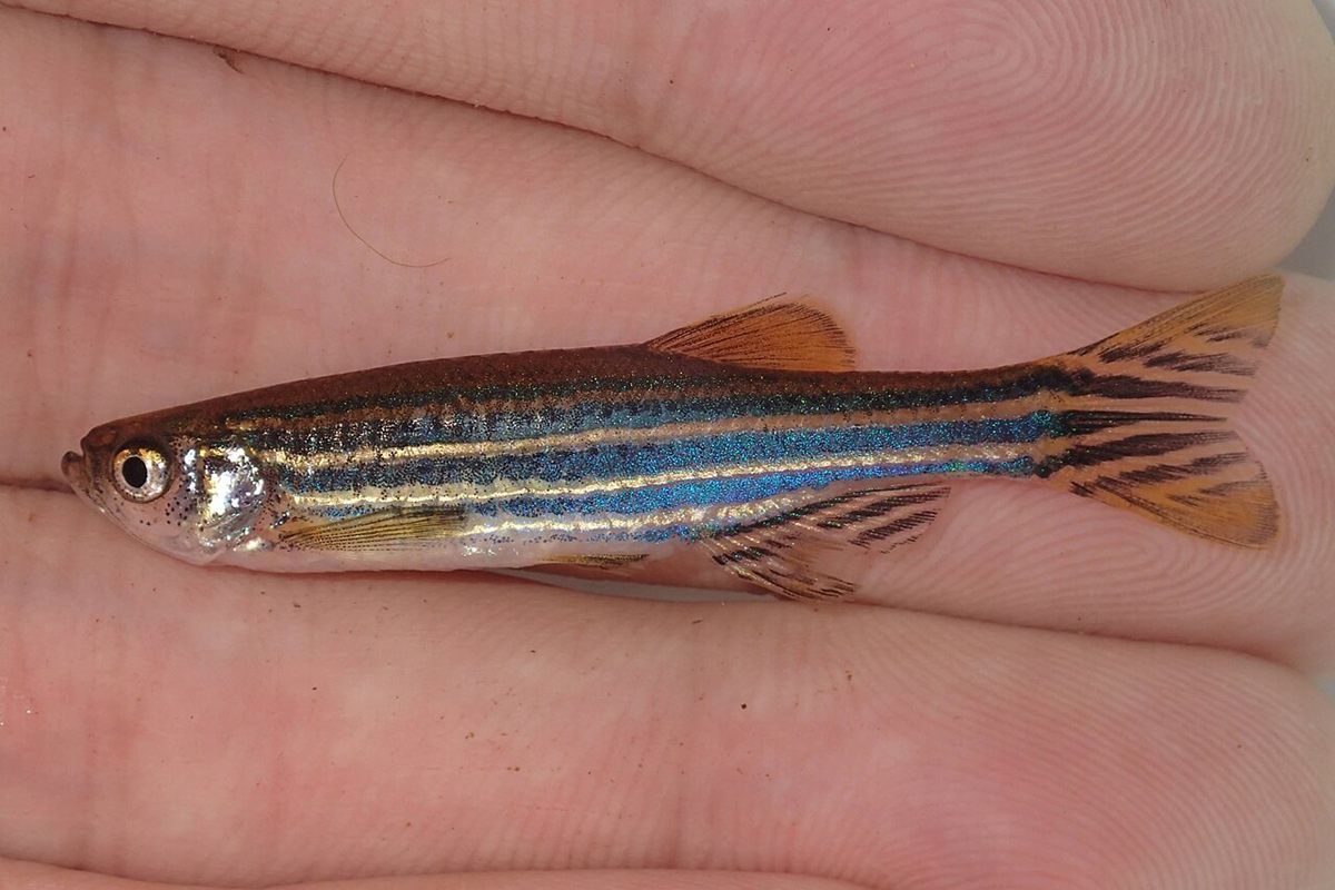
The zebrafish (Danio rerio) is a small striped freshwater fish from South Asia that is one of the most widely used vertebrate model organisms. Its embryos are transparent and develop outside the mother, so researchers can watch organs form in a living animal.
The zebrafish (Danio rerio) is a small striped freshwater fish from South Asia that is one of the most widely used vertebrate model organisms. Its embryos are transparent and develop outside the mother, so researchers can watch organs form in a living animal.
🧬 Field Guide Specimen: Open Zebrafish in the CRV Science Field Guide to log the specimen, see its place among the Vertebrates lineages in the Cladogram Explorer, and test your recall in the Red List conservation game.
Zebrafish at a Glance
| Property | Value |
|---|---|
| Scientific name | Danio rerio |
| Kingdom | Animalia |
| Phylum | Chordata |
| Class | Actinopterygii |
| Infraclass | Teleostei |
| Field Guide group | Marine and Freshwater Fish |
| Diagnostic feature (Field Guide) | Powerful vertebrate biomedical model; embryos are completely transparent. |
| IUCN Red List status | Least Concern (LC), assessed 2010 |
| Genome | About 70% of human genes have a counterpart |
| Native range | South Asia |
Evolutionary Systematics & Comparative Anatomy
Danio rerio (Hamilton, 1822)—originally described as Cyprinus rerio by Francis Buchanan-Hamilton from the Kosi River basin of northern India and long cited in twentieth-century experimental literature under the subgeneric synonym Brachydanio rerio—is a freshwater ray-finned fish belonging to the family Danionidae (formerly classified as subfamily Danioninae within Cyprinidae) and order Cypriniformes (Superorder Ostariophysi, Infraclass Teleostei). Within the genus Danio, which comprises more than 25 small species native to South and Southeast Asia, phylogenomic analyses place D. rerio as sister to Danio aesculapii and Danio kyathit, nested within a clade that exhibits striking evolutionary diversity in pigment pattern organization (ranging from horizontal stripes to vertical bars and uniform spots).
Like all core teleosts, the ancestral lineage of Danio rerio underwent the teleost-specific third-round whole-genome duplication (Ts3R) approximately 320 to 350 million years ago, followed by differential ohnolog retention and subfunctionalization. The haploid nuclear genome of Danio rerio spans ~1.41 gigabases organized across 25 chromosomes (2n = 50) and encodes 26,206 protein-coding genes. High-quality reference assembly comparisons (GRCz11) demonstrate that 71.4% of human protein-coding genes possess at least one zebrafish ortholog, rising to 82% among genes listed in the Online Mendelian Inheritance in Man (OMIM) database as implicated in human genetic pathology.
Pigment Chromatophores, Weberian Apparatus, and Pharyngeal Jaws
Adult Danio rerio measure 3.0 to 4.5 cm in standard length, featuring a fusiform, laterally compressed body, a superior (upturned) lower jaw adapted for surface feeding, and two pairs of sensory barbels—shorter rostral barbels and longer maxillary barbels—supplied by facial nerve (nVII) taste buds. The lateral flank pattern consists of four to five dark blue-black longitudinal stripes alternating with bright silvery-gold interstripes extending from behind the operculum to the caudal fin rays. Cellularly, this Turing-type reaction-diffusion pattern is constructed via short- and long-range contact interactions among three neural-crest-derived dermal chromatophore populations: black melanin-containing melanophores, yellow/orange pteridine- and carotenoid-containing xanthophores, and reflective guanine-platelet iridophores. Engineered double-mutant laboratory strains such as casper—which combines the mitfaw2 mutation (nacre, lacking melanophores) with the mpv17a9 mutation (roy orbison, lacking iridophores)—remain optically translucent throughout adulthood, permitting non-invasive in vivo fluorescence microscopy of internal organs and metastatic cell migration.
As an ostariophysan teleost, D. rerio possesses a Weberian apparatus, a chain of four modified anterior vertebral ossicles (the claustrum, scaphium, intercalarium, and tripus) derived from the first four vertebrae that mechanically couples the anterior chamber of the two-chambered physostomous swim bladder to the perilymphatic sinus of the inner ear, amplifying acoustic pressure oscillations across frequencies from 100 to 4,000 Hz. Whereas the oral jaws lack teeth, the fifth ceratobranchial arch in the posterior pharynx is enlarged into a tooth-bearing pharyngeal jaw armed with two rows of hooked, replaceable pharyngeal teeth that grind ingested crustaceans and insects against a keratinized basioccipital chewing pad on the ventral neurocranium.
Physiology, Biomechanics & Sensory Biology
Ex Utero Embryogenesis and High-Throughput Genetic Tractability
Pioneered as a vertebrate developmental genetics model by George Streisinger at the University of Oregon during the late 1960s and 1970s and scaled to saturation forward-genetic mutagenesis screens by Christiane Nüsslein-Volhard and Wolfgang Driever in the 1990s, Danio rerio bridges the experimental gap between invertebrate genetic models (Drosophila melanogaster, Caenorhabditis elegans) and mammalian systems (Mus musculus). Following external fertilization at 28.5 °C, the telolecithal egg undergoes synchronous meroblastic discoidal cleavage atop the yolk cell at 15-minute intervals until the mid-blastula transition (cell cycle 10, ~3 hours post-fertilization, hpf), when zygotic genome activation and epiboly commence.
Because the chorion and embryonic tissues lack heavy pigmentation during the first 24 hpf (and melanogenesis can be blocked pharmacologically with 1-phenyl-2-thiourea), every mitotic division, gastrulation convergence-extension movement, and neural crest migration can be imaged at single-cell resolution by differential interference contrast or light-sheet microscopy. Somite segmentation begins at 10 hpf, the linear heart tube initiates peristaltic contractions at 22–24 hpf, blood erythrocytes circulate through intersegmental vessels by 26 hpf, and the larva hatches from its chorion between 48 and 72 hpf with a functional visual system, lateral line neuromasts, hepatic and pancreatic primordia, and free-swimming locomotor circuitry by 5 days post-fertilization (dpf). Whole-brain functional calcium imaging in 5–7 dpf transgenic larvae expressing genetically encoded calcium indicators (elavl3:GCaMP6s) allows simultaneous recording of activity across all ~100,000 neurons in the vertebrate brain during optomotor and hunting behaviors.
Epimorphic Cardiac, Retinal, and Spinal Cord Regeneration
Unlike adult mammals, whose injured myocardium heals by fibrotic collagen scar deposition due to cardiomyocyte cell-cycle arrest, adult Danio rerio regenerate up to 20% of their two-chambered heart ventricle within 30 to 60 days following surgical apical resection or cryoinjury without persistent scarring. Lineage-tracing experiments demonstrate that cardiac regeneration does not depend on undifferentiated stem cells; instead, surviving differentiated cardiomyocytes adjacent to the wound border dedifferentiate, disassemble their sarcomeric contractile arrays, upregulate gata4 and cell-cycle regulators (such as Polo-like kinase 1, plk1), and proliferate to replace the lost myocardium, guided by epicardial retinoic acid signaling and transient extracellular matrix fibronectin synthesis. Analogously, after retinal phototoxic or mechanical ablation, resident Müller glia reprogram via ascl1a and lin28 activation into multipotent retinal progenitors that regenerate all neuronal classes (photoreceptors, bipolar, amacrine, and ganglion cells), while transected adult spinal cords bridge glial lesions via fibroblast growth factor (FGF)-driven axonal regrowth and ependymo-radial glial neurogenesis.
Trophic Ecology, Life History & Reproduction
Floodplain Microhabitat, Omnivorous Foraging, and Shoaling
In its native range across the Ganges, Brahmaputra, and Indus river basins of India, Bangladesh, Nepal, and Myanmar, wild Danio rerio inhabits shallow, slow-moving or standing floodplain margins, oxbow ponds, vegetated irrigation ditches, and flooded rice paddies characterized by silt substrates, seasonal monsoon inundation, and wide thermal fluctuations (16 to 38 °C). Wild individuals form cohesive, mixed-sex shoals of 5 to 30+ fish near the water surface, where they feed omnivorously on planktonic crustaceans (cladocerans, copepods, ostracods), aquatic insect larvae (chironomids and culicid mosquito larvae), terrestrial insects fallen onto the surface film, filamentous algae, and vascular plant detritus. When threatened by piscivorous snakeheads (Channa spp.), needlefishes (Xenentodon cancila), or kingfishers, damaged epidermal club cells release an alarm substance (Schreckstoff, containing hypoxanthine-3-N-oxide and chondroitin fragments) that triggers immediate shoal cohesion, rapid erratic dashing, and substrate diving in conspecifics via olfactory receptor neurons.
Photoperiodic Spawning and Polygenic Sex Determination
In nature, reproductive maturation is coupled to the onset of the Southwest monsoon (May to August), when warm rainfall dilutes water conductivity and floods vegetated shallow margins, whereas domesticated laboratory strains spawn year-round at dawn when photoperiod lights switch on. Courtship involves rapid pursuit in which the male nudges the female's vent and encircles her over gravel or aquatic vegetation, stimulating the release and external fertilization of 100 to 300 non-adhesive demersal eggs (0.7 mm diameter) per clutch without subsequent parental care. Wild Danio rerio strains possess a genetic sex determination system centered on a dominant female-determining locus on the right arm of chromosome 4 (ZW/ZZ), whereas domesticated laboratory strains (such as AB and Tübingen) lost the chromosome 4 sex determinant during artificial selection and rely on a polygenic sex determination mechanism modulated by juvenile primordial germ cell numbers, temperature, and growth rate: individuals maintain a bipotential juvenile ovary until 20–25 dpf, after which persistence of high oocyte numbers stabilizes female differentiation via aromatase (cyp19a1a) estrogen synthesis, whereas oocyte apoptosis triggers testicular differentiation.
Frequently Asked Questions
Why did biologists choose the zebrafish as a model for vertebrate development and human disease?
Unlike mice, whose embryos develop inside an opaque maternal uterus, zebrafish females release hundreds of externally fertilized eggs that remain optically transparent throughout organ formation. Combined with rapid development—producing a beating heart and circulating blood within 24 hours—and an assembled genome sharing orthologs with over 70% of human genes, zebrafish allow live microscopic tracking of genetic mutations and drug effects.
How does an adult zebrafish regenerate its heart muscle after injury?
When up to one-fifth of the zebrafish heart ventricle is surgically removed or damaged, the wound seals with a temporary fibrin clot that is gradually replaced by functional muscle rather than permanent scar tissue. Existing heart muscle cells (cardiomyocytes) near the injury border partially dedifferentiate, dismantle their contractile fibers, re-enter mitosis, and divide to rebuild the myocardium.
What is the casper zebrafish strain and how is it used in research?
While wild-type zebrafish embryos are transparent for their first few days, adult zebrafish develop opaque dark stripes of melanin and reflective guanine crystals that block deep-tissue microscopy. The engineered casper strain carries double mutations (nacre and roy orbison) that eliminate both black melanophores and reflective iridophores, leaving the adult fish translucent so scientists can track transplanted stem cells or tumors live.
References
- Howe, K., Clark, M. D., Torroja, C. F., Torrance, J., Berthelot, C., Muffato, M., Collins, J. E., Humphray, S., McLaren, K., Matthews, L., McLaren, S., Sealy, I., Caccamo, M., Churcher, C., Scott, C., Barrett, J. C., Koch, R., Rauch, G.-J., White, S., ... Stemple, D. L. (2013). The zebrafish reference genome sequence and its relationship to the human genome. Nature, 496(7446), 498–503. https://doi.org/10.1038/nature12111
- Poss, K. D., Wilson, L. G., & Keating, M. T. (2002). Heart regeneration in zebrafish. Science, 298(5601), 2188–2190. https://doi.org/10.1126/science.1077857
- Jopling, C., Sleep, E., Raya, M., Martí, M., Raya, A., & Izpisúa Belmonte, J. C. (2010). Zebrafish heart regeneration occurs by cardiomyocyte dedifferentiation and proliferation. Nature, 464(7288), 606–609. https://doi.org/10.1038/nature08899
- White, R. M., Sessa, A., Burke, C., Bowman, T., LeBlanc, J., Ceol, C., Bourque, C., Dovey, M., Goessling, W., Burns, C. E., & Zon, L. I. (2008). Transparent adult zebrafish as a tool for in vivo transplantation analysis. Cell Stem Cell, 2(2), 183–189. https://doi.org/10.1016/j.stem.2007.11.002