American Dog Tick
The American dog tick (Dermacentor variabilis) is a common tick of eastern and central North America. It is the main vector of Rocky Mountain spotted fever in the eastern United States and of tularemia.
The American dog tick (Dermacentor variabilis) is a common tick of eastern and central North America. It is the main vector of Rocky Mountain spotted fever in the eastern United States and of tularemia.
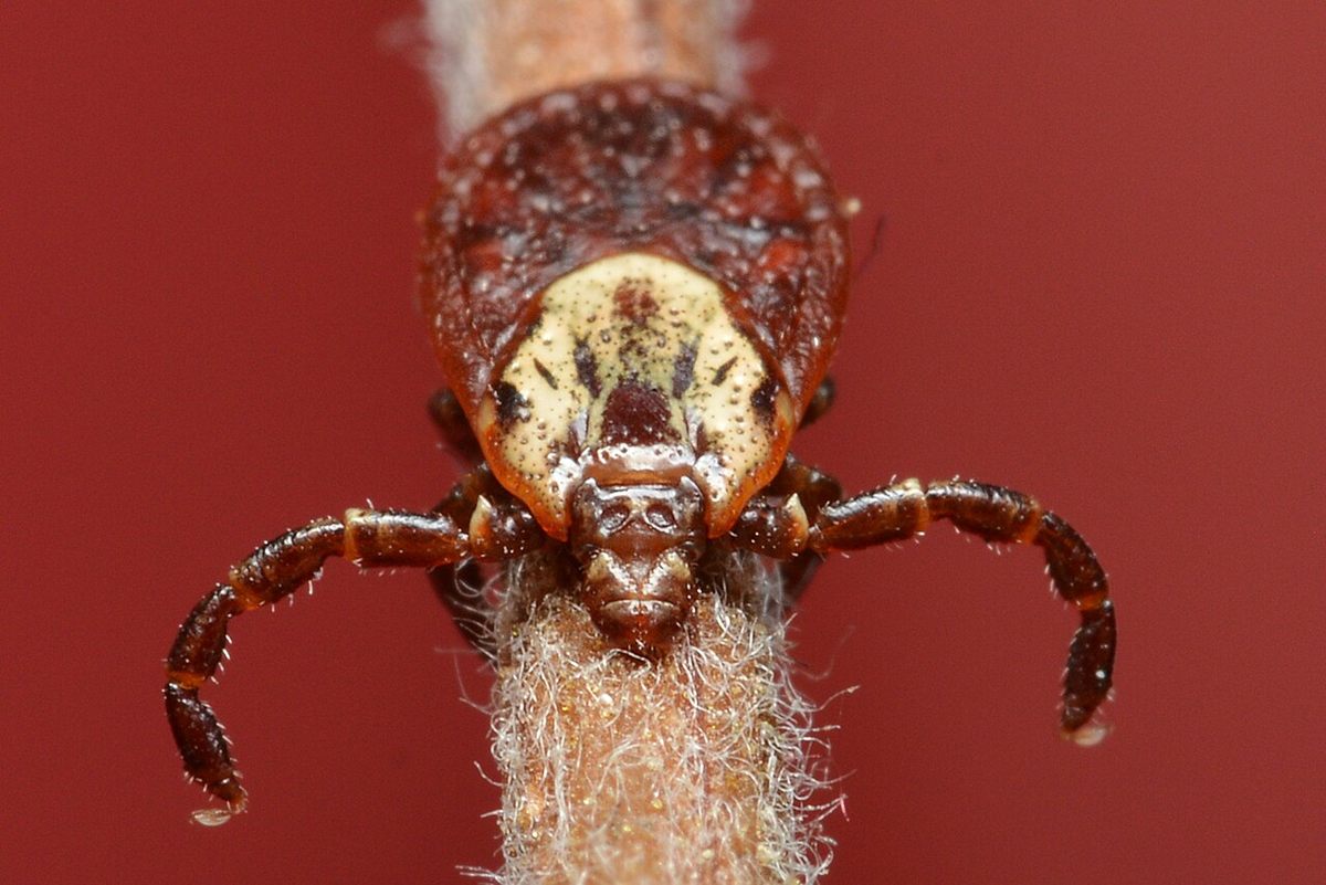
🧬 Field Guide Specimen: Open American Dog Tick in the CRV Science Field Guide to log the specimen, see its place among the Ecdysozoa lineages in the Cladogram Explorer, and test your recall in the Red List conservation game.
American Dog Tick at a Glance
| Property | Value |
|---|---|
| Scientific name | Dermacentor variabilis |
| Kingdom | Animalia |
| Phylum | Arthropoda |
| Class | Arachnida |
| Order | Ixodida |
| Family | Ixodidae |
| Field Guide group | Ecdysozoa: Arachnida (Spiders, Scorpions & Allies) |
| Diagnostic feature (Field Guide) | Brown eight-legged blood-feeder with an ornate cream-marbled shield; waits on grass tips to grab passing dogs and people. |
| Order | Ixodida |
| Disease | Rocky Mountain spotted fever |
Taxonomy, Metastriate Phylogenetics, and Cryptic Western Divergence
The American dog tick (Dermacentor variabilis, also known as the wood tick) is an obligate hematophagous ectoparasite belonging to the arachnid subclass Acari, superorder Parasitiformes, order Ixodida, and hard-tick family Ixodidae. Within Ixodidae, ticks are divided into two major evolutionary lineages based on the position of the anal groove relative to the anus: the basal Prostriata, comprising the single genus Ixodes (which includes the North American Blacklegged Tick, Ixodes scapularis, and the European Castor Bean Tick, Ixodes ricinus), and the derived Metastriata, which includes the genera Dermacentor, Amblyomma, Rhipicephalus, and Haemaphysalis. Unlike prostriate Ixodes ticks, which lack eyes and festoons and possess an anal groove arching anterior to the anus, metastriate Dermacentor ticks possess a posterior anal groove, eleven rectangular marginal festoons along the posterior idiosoma, laterally positioned simple eyes on the scutum, and an "ornate" dorsal shield patterned with iridescent ivory-white cuticular enamel over a dark reddish-brown base.
East–West Phylogeography and Dermacentor similis
Across North America, Dermacentor variabilis sensu stricto is distributed broadly from Atlantic Canada (Nova Scotia) and the eastern United States westward to the Great Plains and south into northeastern Mexico. For decades, disjunct populations along the Pacific Coast (California, Oregon, and Washington) were treated as a western population of D. variabilis. In 2021, integrative genomic and morphological revisions demonstrated that these western coastal populations had been reproductively isolated from eastern D. variabilis by the Rocky Mountains for millions of years, resulting in the formal description of the western dog tick (Dermacentor similis). Meanwhile, in the higher elevations of the Rocky Mountain cordillera, the sister species Dermacentor andersoni (the Rocky Mountain wood tick) replaces D. variabilis ecologically.
Gnathosomal Anatomy, Three-Host Life Cycle, and Salivary Osmoregulation
Like all ixodid hard ticks, Dermacentor variabilis exhibits an obligate three-host life cycle spanning one to three years depending on microclimate and host availability. Gravid females drop off their final vertebrate host into leaf litter, oviposit a single glutinous mass of 4,000 to 6,500 eggs over two to four weeks, and die. Six-legged larvae ("seed ticks") hatch and quest low in the herbaceous boundary layer to attach to small terrestrial rodents—primarily meadow voles (Microtus pennsylvanicus), white-footed mice (Peromyscus leucopus), and cotton rats (Sigmodon hispidus)—feeding for 3 to 5 days before dropping to the ground to molt into eight-legged nymphs. Nymphs similarly quest for small- to medium-sized mammals (voles, mice, cottontail rabbits, raccoons, and opossums), feed for 4 to 7 days, and drop off to molt into sexually dimorphic adults. In adult males, the ornate dorsal scutum covers the entire dorsal surface of the idiosoma, restricting abdominal expansion; in adult females, the scutum is restricted to the anterior third of the dorsum, allowing the soft, pleated alloscutum cuticle to synthesize new endocuticle and expand during a 7-to-12-day blood meal on medium-to-large mammals (domestic dogs, coyotes, foxes, white-tailed deer, and humans), increasing the female's body mass more than 100-fold (from ~5 mg unfed to 500–700 mg engorged).
Haller's Organ, Cement Cone Attachment, and Salivary Fluid Secretion
To locate hosts along forest edges and old-field trails, adult D. variabilis climb grass stems to heights of 30 to 90 cm and adopt a "questing" posture with their forelegs extended. The dorsal tarsus of each first walking leg houses Haller's organ—a complex sensory pit and capsule containing multiporous olfactory, thermoreceptive, and hygroreceptive sensilla that detect host-emitted CO2 plumes, butyric acid, ammonia, radiant infrared heat, and substrate vibrations. Once aboard a host, the tick uses its paired telescoping chelicerae to lacerate the epidermis and inserts its barbed, median ventral hypostome into the dermal capillary pool. Within hours of attachment, the tick's acinar salivary glands secrete a glycine- and tyrosine-rich proteinaceous cement that polymerizes around the mouthparts into an anchor cone. Because blood is ~80% water, an engorging female concentrates the ingested erythrocytes and plasma proteins in her midgut diverticula and pumps roughly 65–75% of the ingested water and excess Na+ and Cl− ions back into the host bloodstream via dopamine-stimulated type II and III salivary acini, simultaneously injecting vasodilators, platelet aggregation inhibitors, and immunomodulatory proteins (such as evasins that bind host chemokines).
| Life Stage / Anatomical Structure | Morphological & Physiological Characteristics | Vector / Ecological Function |
|---|---|---|
| Six-legged larva & eight-legged nymph | Microscopic stages (0.6 mm and 1.5 mm) lacking genital aperture; quest < 15 cm in litter | Feed on Microtus voles and Peromyscus mice; amplify enzootic Rickettsia and Francisella |
| Adult female & male | 3–5 mm unfed (15 mm engorged female); short basis capituli; ornate white-marbled scutum | Quest on tall grass stems (30–90 cm); mate on large mammalian hosts; bridge pathogens to dogs and humans |
| Haller's organ (Foreleg tarsus I) | Anterior sensory pit and posterior capsule housing chemosensory and thermal sensilla | Detects host CO2, NH3, lactic acid, and radiant body heat |
| Salivary cement & osmoregulation | Glycine-rich cement cone + dopamine-activated Na+/K+-ATPase salivary fluid return | Locks short mouthparts in dermis for 7–12 days and returns ~70% of blood-meal water into the host |
| Vector refractoriness to Borrelia | Hemolymph defensins and midgut proteases rapidly lyse ingested Borrelia burgdorferi | Explains why D. variabilis is completely incompetent to transmit Lyme disease spirochetes |
Rickettsial Pathogenesis, Tularemia, and Ascending Tick Paralysis
In human and veterinary medicine, Dermacentor variabilis is the primary vector of Rocky Mountain spotted fever (RMSF) across the eastern, central, and southern United States, where the majority of U.S. clinical cases occur. The causative agent, Rickettsia rickettsii, is an obligate intracellular alpha-proteobacterium (see Bacteria) maintained in nature through both horizontal amplification in small rodent reservoirs and transovarial and transstadial transmission inside the tick. When an infected tick attaches to a vertebrate host, dormant rickettsiae in the salivary glands require a 6-to-20-hour "reactivation" phase triggered by the warm blood meal and physiological temperature (37 °C) to restore virulence before entering the host dermis. Once inoculated, R. rickettsii invades vascular endothelial cells lining small arteries, capillaries, and venules across the body, polymerizing host actin filaments to propel itself from cell to cell. Endothelial injury leads to widespread microvascular leakage, petechial rash (classically beginning on the wrists and ankles and spreading to the palms and soles), thrombocytopenia, hyponatremia, cerebral edema, and non-cardiogenic pulmonary edema, carrying a 20–25% case-fatality rate if left untreated, though rapid oral or intravenous doxycycline therapy arrests bacterial replication.
Francisella tularensis, Canine Pathogens, and Neurotoxic Tick Paralysis
Beyond RMSF, Dermacentor variabilis transmits Francisella tularensis (the Gram-negative coccobacillus causing ulceroglandular tularemia), Ehrlichia canis (canine monocytic ehrlichiosis), Anaplasma marginale (bovine anaplasmosis), and Cytauxzoon felis (an apicomplexan hemoprotozoan causing fatal cytauxzoonosis in domestic cats). In addition to infectious pathogens, a single engorging adult female D. variabilis can induce acute, non-infectious tick paralysis in humans (especially children) and dogs. After 5 to 7 days of continuous feeding, the female's salivary glands secrete a neurotoxic holocyclotoxin-like peptide that inhibits acetylcholine release at motor nerve terminals and slows nerve conduction velocity, producing an acute, symmetrical, ascending flaccid paralysis with areflexia that closely mimics Guillain–Barré syndrome and can paralyze the respiratory diaphragm. Unlike infectious neuropathies or botulism, complete mechanical removal of the attached tick (including the capitulum) results in dramatic neurological recovery within 12 to 24 hours. Conversely, despite frequently feeding on Borrelia burgdorferi-infected white-footed mice, Dermacentor variabilis cannot transmit Lyme disease because antimicrobial peptides (specifically defensin varisin) and complement-like factors in its midgut and hemolymph rapidly lyse ingested Borrelia spirochetes before they can colonize the salivary glands.
Frequently Asked Questions
Why can't the American dog tick transmit Lyme disease like the blacklegged tick?
When an American dog tick (Dermacentor variabilis) feeds on a mouse infected with the Lyme disease spirochete (Borrelia burgdorferi), the tick ingests the bacteria into its midgut. However, unlike the blacklegged tick (Ixodes scapularis), the American dog tick lacks the midgut receptor protein (TROSPA) that Borrelia needs to anchor itself, and the dog tick's immune system secretes potent antimicrobial defensin peptides that rapidly rupture and kill the spirochetes within hours of ingestion.
Why do most cases of "Rocky Mountain" spotted fever occur in the eastern and southeastern United States?
Rocky Mountain spotted fever was first identified in the 1890s in the Bitterroot Valley of Montana, where it is transmitted by the Rocky Mountain wood tick (Dermacentor andersoni). However, the same intracellular bacterium (Rickettsia rickettsii) is also transmitted throughout the eastern, central, and southeastern United States by the far more abundant and suburban-adapted American dog tick (Dermacentor variabilis), making states such as North Carolina, Tennessee, Oklahoma, Arkansas, and Missouri the primary geographic centers of the disease.
How does tick paralysis happen without any bacterial or viral infection?
Tick paralysis is a direct toxic envenomation rather than an infectious disease. As a gravid female American dog tick enters the rapid engorgement phase after 5 to 7 days of feeding, her salivary glands secrete a neurotoxin into the host's bloodstream that blocks acetylcholine release at neuromuscular junctions. This causes a progressive paralysis that starts in the legs and ascends toward the chest; simply finding and removing the feeding tick halts toxin delivery and reverses the paralysis within hours.
References
- Sonenshine, D. E., & Roe, R. M. (Eds.). (2014). Biology of Ticks (2nd ed., Vols. 1–2). Oxford University Press. https://doi.org/10.1093/oso/9780199744053.001.0001
- Lado, P., Glon, M. G., Klompen, H., & Short, M. (2021). Integrative taxonomy of Dermacentor variabilis (Ixodida: Ixodidae) with description of a new species, Dermacentor similis n. sp. Journal of Medical Entomology, 58(6), 2216–2227. https://doi.org/10.1093/jme/tjab134
- Johns, R., Sonenshine, D. E., & Hynes, W. L. (2001). Identification of a defensin from the hemolymph of the American dog tick, Dermacentor variabilis. Insect Biochemistry and Molecular Biology, 31(9), 857–865. https://doi.org/10.1016/S0965-1748(01)00031-500031-5)
- Biggs, H. M., Behravesh, C. B., Bradley, K. K., Dahlgren, F. S., Drexler, N. A., Dumler, J. S., Folk, S. M., Kato, C. Y., Lash, R. R., Levin, M. L., Massung, R. F., Nadelman, R. B., Nicholson, W. L., Paddock, C. D., Pritt, B. S., & Traeger, M. S. (2016). Diagnosis and management of tickborne rickettsial diseases: Rocky Mountain spotted fever and other spotted fever group rickettsioses, ehrlichioses, and anaplasmosis — United States. MMWR Recommendations and Reports, 65(2), 1–44. https://doi.org/10.15585/mmwr.rr6502a1