Blacklegged Tick
The blacklegged tick or deer tick (Ixodes scapularis) is the main vector of Lyme disease in eastern and central North America. It also transmits anaplasmosis, babesiosis and Powassan virus, and its range is expanding northward.
The blacklegged tick or deer tick (Ixodes scapularis) is the main vector of Lyme disease in eastern and central North America. It also transmits anaplasmosis, babesiosis and Powassan virus, and its range is expanding northward.
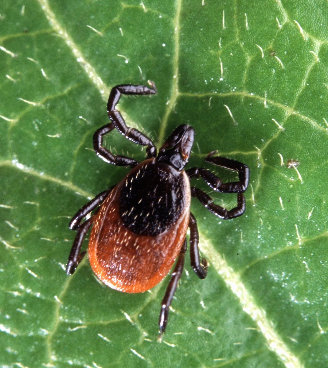
🧬 Field Guide Specimen: Open Blacklegged Deer Tick in the CRV Science Field Guide to log the specimen, see its place among the Ecdysozoa lineages in the Cladogram Explorer, and test your recall in the Red List conservation game.
Blacklegged Tick at a Glance
| Property | Value |
|---|---|
| Scientific name | Ixodes scapularis |
| Kingdom | Animalia |
| Phylum | Arthropoda |
| Subphylum | Chelicerata |
| Class | Arachnida |
| Order | Ixodida |
| Field Guide group | Ecdysozoa: Arachnida (Spiders, Scorpions & Allies) |
| Diagnostic feature (Field Guide) | Dorsoventrally flattened leathery unsegmented body (idiosoma) and barbed anterior hypostome mouthparts for anchoring. |
| Order | Ixodida |
| Pathogens carried | Borrelia burgdorferi and others |
Taxonomy, Phylogenetics, and Evolutionary History
Described by Thomas Say in 1821, the blacklegged tick (Ixodes scapularis) belongs to the prostriate hard-tick family Ixodidae (order Ixodida, class Arachnida). Within Ixodes, it is a member of the Holarctic Ixodes ricinus species complex alongside the western blacklegged tick (I. pacificus), the European sheep tick (I. ricinus), and the Eurasian taiga tick (I. persulcatus).
In 1979, Andrew Spielman and colleagues erected the name Ixodes dammini for northern deer ticks driving the New England Lyme epidemic. Hybridization and DNA analyses by James H. Oliver Jr. and colleagues in 1993 showed that northern and southern populations are conspecific, sinking I. dammini into I. scapularis. Population genomics confirms two clades: the northern clade (whose nymphs quest on exposed leaf litter and twigs, driving high human Lyme incidence) and the southern clade (whose nymphs parasitize skinks beneath the litter). Its 2.1-gigabase genome (2016) reveals expansions of salivary Kunitz protease inhibitors and lipocalins.
Comparative Anatomy, Haller's Organ, and Salivary Immunomodulation
Gnathosoma Mechanics and Haller's Organ Chemoreception
Ixodes scapularis divides its body into an anterior gnathosoma (mouthparts) and a posterior unsegmented idiosoma bearing the legs (six in larvae; eight in nymphs and adults) and dorsal scutum. In adult males (~2.0–2.5 mm), the dark holoscutum covers the dorsum; in adult females (~3.0–3.5 mm unfed, with an orange-red posterior alloscutum), extensible endocuticle allows engorgement to 10 millimeters and an 80- to 100-fold mass gain over 5 to 7 days.
Lacking eyes, I. scapularis detects hosts via Haller's organ on tarsus I of the forelegs, sensing carbon dioxide (CO2), ammonia, lactic and butyric acids, and infrared heat (37 °C). Paired chelicerae incise the skin to insert the barbed median hypostome, anchored by a salivary cement cone.
| Anatomical or Salivary Component | Molecular / Structural Identity | Physiological or Pathogenic Function |
|---|---|---|
| Haller's organ on tarsus I | Sensory pit housing olfactory, hygro-, and thermoreceptive sensilla | Detects host CO2 plumes, butyric acid, and 37 °C infrared radiation |
| Recurved hypostome & cement | Barbed median subcheliceral anchor and cross-linked salivary cement proteins | Locks mouthparts into host dermis during 3- to 7-day blood meal |
| Salp15 salivary glycoprotein | 15-kDa immunosuppressive protein binding host CD4+ T-cells and OspC | Inhibits host IL-2 production and shields Borrelia burgdorferi from complement |
| Ixolaris & Salp14 anticoagulants | Tissue factor pathway inhibitor (TFPI) and Factor Xa inhibitors in saliva | Prevents blood coagulation in the dermal hemorrhagic feeding pool |
| OspA-to-OspC spirochete switch | Temperature- and blood-pH-triggered surface lipoprotein switch in B. burgdorferi | Detaches spirochetes from tick midgut TROSPA receptor for salivary migration |
Salivary Pharmaco-Immunology and the OspA-to-OspC Transmission Delay
Unlike capillary-feeding mosquitoes (see Mosquito-borne disease), I. scapularis is a telmophage (pool feeder) that lacerates microvessels and injects anticoagulants (ixolaris, Salp14), histamine-binding lipocalins, complement inhibitors (Isac, Salp20), and Salp15 (blocking CD4+ T-cell IL-2 transcription). In the unfed midgut, Borrelia burgdorferi (see Bacteria) expresses OspA, binding the midgut receptor TROSPA. When warm (37 °C) blood enters the gut, the RpoN–RpoS cascade downregulates ospA and upregulates OspC, releasing spirochetes to migrate to the salivary glands, bind Salp15, and enter the dermis 36 to 48 hours after attachment.
Two-Year Three-Host Life Cycle, Multi-Pathogen Ecology, and Range Expansion
Across eastern and north-central North America, I. scapularis completes a two-year, three-host life cycle:
- Year 1 (Eggs and Larvae): Females lay 1,500 to 3,000 eggs in May. Six-legged larvae hatch in July–August free of B. burgdorferi and feed once on small vertebrates—chiefly the white-footed mouse (Peromyscus leucopus)—acquiring B. burgdorferi before molting into overwintering nymphs.
- Year 2 Spring–Summer (Nymphs): Infected nymphs (~1.5 mm) quest in May–July, infecting juvenile mice prior to August larval hatching and causing over 80% of human Lyme cases.
- Year 2 Autumn–Year 3 Spring (Adults): Adults feed and mate on white-tailed deer (Odocoileus virginianus), whose serum complement lyses B. burgdorferi but whose blood sustains tick reproduction.
Alongside B. burgdorferi (treated with doxycycline), I. scapularis transmits Borrelia miyamotoi, Anaplasma phagocytophilum, Babesia microti, and salivary Powassan virus (transmitted in 15 minutes). Driven by reforestation and Climate change, its range is expanding into Canada.
Frequently Asked Questions
Why does a blacklegged tick usually need to stay attached for 36 to 48 hours to transmit Lyme disease, whereas Powassan virus can be transmitted in 15 minutes?
In an unfed tick, Borrelia burgdorferi is anchored to the midgut wall by Outer Surface Protein A (OspA). Only after warm blood enters the gut does the bacterium switch from OspA to OspC, detach, and migrate to the salivary glands over 36 to 48 hours. Conversely, Powassan virus already resides in the salivary glands of an unfed tick and enters the bite wound within 15 minutes.
Why are white-footed mice the primary reservoir for Lyme disease while white-tailed deer do not infect ticks with Borrelia burgdorferi?
White-footed mice (Peromyscus leucopus) tolerate high levels of Borrelia burgdorferi in their skin and blood, infecting 75% to 90% of feeding larval ticks. Conversely, white-tailed deer (Odocoileus virginianus) possess innate serum complement proteins that lyse B. burgdorferi on contact; instead, deer drive the cycle indirectly as the primary reproductive host for adult female ticks.
Why do poppy-seed-sized nymphs in late spring cause far more human Lyme disease cases than larger adult ticks in autumn?
Although adult females in autumn have a higher infection rate (~50%) than nymphs (~20%–25%), adults are larger, easier to remove within 24 hours, and quest when people wear cold-weather clothing. Nymphs quest in May–July when people wear summer clothing, and their 1.5-millimeter size and analgesic saliva allow them to feed undetected for 48 hours.
References
- Spielman, A., Wilson, M. L., Levine, J. F., & Piesman, J. (1985). Ecology of Ixodes dammini-borne human babesiosis and Lyme disease. Annual Review of Entomology, 30(1), 439–460. https://doi.org/10.1146/annurev.en.30.010185.002255
- Schwan, T. G., Piesman, J., Golde, W. T., Dolan, M. C., & Rosa, P. A. (1995). Induction of an outer surface protein on Borrelia burgdorferi during tick feeding. Proceedings of the National Academy of Sciences, 92(7), 2909–2913. https://doi.org/10.1073/pnas.92.7.2909
- Ramamoorthi, N., Narasimhan, S., Pal, U., Bao, F., Yang, X. F., Fish, D., Anguita, J., Norgard, M. V., Kantor, F. S., Anderson, J. F., Koski, R. A., & Fikrig, E. (2005). The Lyme disease agent exploits a tick protein to infect the mammalian host. Nature, 436(7050), 573–577. https://doi.org/10.1038/nature03812
- Gulia-Nuss, M., Nuss, A. B., Meyer, J. M., Sonenshine, D. E., Roe, R. M., Waterhouse, R. M., Sattelle, D. B., de la Fuente, J., Ribeiro, J. M., Megy, K., Thimmapuram, J., Miller, J. R., et al. (2016). Genomic insights into the Ixodes scapularis tick vector of Lyme disease. Nature Communications, 7, 10507. https://doi.org/10.1038/ncomms10507