Melon Comb Jelly
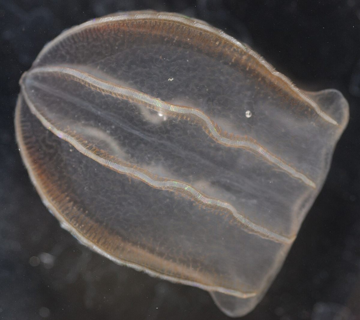
The melon comb jelly (Beroe ovata) is a pinkish, melon-shaped comb jelly with a huge mouth and no tentacles. It feeds almost exclusively on other comb jellies.
The melon comb jelly (Beroe ovata) is a pinkish, melon-shaped comb jelly with a huge mouth and no tentacles. It feeds almost exclusively on other comb jellies.
🧬 Field Guide Specimen: Open Melon Comb Jelly in the CRV Science Field Guide to log the specimen, see its place among the Cnidaria+Ctenophora lineages in the Cladogram Explorer, and test your recall in the Red List conservation game.
Melon Comb Jelly at a Glance
| Property | Value |
|---|---|
| Scientific name | Beroe ovata |
| Kingdom | Animalia |
| Phylum | Ctenophora |
| Class | Nuda |
| Order | Beroida |
| Family | Beroidae |
| Field Guide group | Cnidaria + Ctenophora (Radiata & Jellies) |
| Diagnostic feature (Field Guide) | Thimble-shaped transparent sac with no tentacles at all; eight rows of shimmering cilia plates run its length, and its huge mouth swallows other jellies whole. |
| Phylum | Ctenophora |
| Diet | Other comb jellies |
Evolutionary Systematics & Comparative Anatomy
Beroe ovata is the type representative of the family Beroidae and order Beroida, the sole order within the class Nuda (or Atentaculata) of the phylum Ctenophora. Whereas all other comb jellies—classified in the class Tentaculata, including cydippids and lobates such as the sea walnut (Mnemiopsis leidyi) and the lobed comb jelly (Bolinopsis infundibulum)—possess paired tentacles armed with glue-secreting colloblasts during at least their cydippid larval stage, members of the class Nuda lack tentacles, tentacle sheaths, and colloblasts throughout their entire ontogeny. Molecular phylogenomic studies of ctenophore genomes and transcriptomes confirm that Ctenophora represents one of the earliest-diverging animal phyla, and indicate that the tentacle-less Beroida evolved secondarily from tentaculate ancestors as specialized macrophagous predators of other gelatinous zooplankton.
Native to the western Atlantic Ocean (from Chesapeake Bay south through the Gulf of Mexico and Caribbean to Argentina) and the Mediterranean Sea, Beroe ovata has a mitriform (thimble- or flattened-melon-shaped), laterally compressed gelatinous body measuring 60 to 160 millimeters in length and 40 to 80 millimeters in width. The transparent mesoglea is tinged milky-pink to reddish-brown in larger adults and traversed by an anastomosing network of meridional gastrovascular canals whose blind-ending lateral branches (diverticula) link across the body wall—a diagnostic anatomical character separating Beroe ovata from the northern Atlantic Beroe cucumis, in which the lateral canal diverticula end blindly without anastomosing.
Nearly the entire internal volume of the animal consists of a cavernous, muscular stomodeum (pharynx) that opens at the oral pole via a wide, slit-like mouth running the full width of the body. At the opposite (aboral) pole sits the apical sensory organ, a dome-shaped statocyst containing a calcareous statolith balanced atop four tufts of mechanosensory balancer cilia, flanked by two branching, papillate polar fields that serve an olfactory-like chemosensory role.
Physiology, Biomechanics & Sensory Biology
Metachronal Ciliary Propulsion & Structural Diffraction
Running longitudinally from the aboral apical organ toward the oral rim are eight meridional comb rows (costae)—four subventral and four subtentacular—each composed of dozens of transverse paddle-like plates called ctenes (comb plates). Each individual ctene measures up to 1 to 2 millimeters wide and consists of thousands of 9+2 cilia fused laterally into a single macrocilium by compartmentalized bridges. Directed by the four balancer tufts of the apical statocyst, the ctenes beat with an oral-to-aboral effective stroke, while the mechanical hydrodynamic wave propagates in the opposite direction (aboral-to-oral antiplectic metachronism) at frequencies of 5 to 20 hertz, propelling Beroe ovata smoothly through the water column at speeds of 1 to 4 centimeters per second.
During daylight, the beating comb rows shimmer with vivid, shifting rainbow colors. This iridescence is not bioluminescence; rather, it is structural light diffraction and interference caused by the periodic sub-micrometer spacing of the thousands of packed ciliary microtubules within each moving ctene acting as a biological diffraction grating. In darkness or when mechanically disturbed, B. ovata also produces true bioluminescence along the eight meridional canals beneath the comb rows, emitting flashes of blue-green light (λmax ≈ 485–490 nanometers) catalyzed by Ca2+-activated berovin photoproteins utilizing coelenterazine as their chromophore, which oxidize to emit a photon without requiring molecular O2.
Reversible Tissue Adhesion & Pharyngeal Macrocilia ("Teeth")
When Beroe ovata searches horizontally or vertically for prey, keeping its wide oral rim open creates excessive hydrodynamic drag. To streamline its anterior profile during cruising flight, the two opposing muscular lips of the mouth zip shut through reversible epithelial cell-to-cell adhesion: interdigitating microvilli and specialized junctional complexes temporarily fuse the upper and lower oral epithelia together so tightly that no mouth opening is visible. When the chemosensory lips contact the mucus or amino acids of another ctenophore, intracellular Ca2+ signaling breaks these adhesive bonds within milliseconds and radial muscular fibers contract, flaring the mouth open into a circular gape wider than the predator's resting body.
Lining the inner oral rim in a dense U-shaped band are thousands of giant, sickle-shaped pharyngeal macrocilia that function as biological teeth. Each macrocilium measures 35 to 50 micrometers in length and 5 to 10 micrometers in diameter, formed by 2,000 to 3,000 individual 9+2 ciliary axonemes bundled within a single shared plasma membrane and cross-linked into a hexagonal paracrystalline lattice powered by dynein arms. When B. ovata encounters a prey comb jelly matchable to its own size, radial pharyngeal dilators generate negative suction pressure to engulf the prey whole; when attacking a much larger lobate ctenophore or salp, the pharyngeal macrocilia beat synchronously inward at 10 to 30 hertz like a chainsaw, rasping through the prey's mesoglea to bite off and swallow discrete chunks of tissue while preventing the slippery gelatinous mass from sliding backward out of the pharynx.
Trophic Ecology, Reproduction & the Black Sea Biological Control
Stenophagous Gelatinous Predation & Simultaneous Hermaphroditism
Beroe ovata is an obligate stenophagous predator that feeds almost entirely on other ctenophores—principally lobate species (Mnemiopsis leidyi, Bolinopsis, and Leucothea) and cydippids (Pleurobrachia pileus). Because the cavernous stomodeum distends up to fourfold during feeding, an individual B. ovata can ingest prey biomass equal to 100 to 300% of its own body weight per day, digesting the gelatinous tissue rapidly within 2 to 5 hours via extracellular proteases in the pharynx followed by intracellular phagocytosis in the meridional canals, and expelling indigestible residues through both the mouth and two aboral anal pores flanking the statocyst.
Like most ctenophores, Beroe ovata is a simultaneous hermaphrodite capable of self-fertilization, with band-like ovaries and testes running side by side along the walls of all eight meridional gastrovascular canals beneath the comb rows. At water temperatures of 20 to 26 °C and high prey availability, adults spawn daily at dusk or dawn, releasing 1,000 to 6,000 gametes through gonopores along the costae. Fertilized eggs (0.8–1.0 millimeter in diameter) undergo biradial ctenophore cleavage and hatch within 24 to 36 hours into miniature, tentacle-less beroid larvae that immediately hunt newly hatched Mnemiopsis and Pleurobrachia larvae, reaching sexual maturity within 12 to 20 days.
Trophic Cascade Recovery in the Black Sea and Caspian Sea
The ecological significance of Beroe ovata was demonstrated during the late-20th-century ecological crisis in the Black Sea. In the early 1980s, the western Atlantic lobate comb jelly sea walnut (Mnemiopsis leidyi) was accidentally introduced into the Black Sea via ship ballast water. Lacking natural predators in the basin and aided by overfishing of planktivorous fishes, M. leidyi proliferated by 1989 to biomasses exceeding 300 to 1,000 grams per square meter (an estimated 800 million metric tons basin-wide). Mnemiopsis devoured the native crustacean zooplankton (copepods and cladocerans) alongside pelagic fish eggs and larvae, causing the collapse of the Black Sea European anchovy (Engraulis encrasicolus) fishery—which plummeted from >600,000 metric tons per year to <100,000 metric tons—while triggering hypoxic phytoplankton blooms.
In autumn 1997, Beroe ovata arrived in the Black Sea (also via ballast water from the western Atlantic or Mediterranean) and established breeding populations by summer 1999. Because B. ovata feeds exclusively on other ctenophores and ignores crustacean zooplankton and fish eggs, its late-summer bloom consumed up to 10 to 60% of the Mnemiopsis leidyi standing stock daily, compressing the annual Mnemiopsis bloom to a brief 4-to-6-week window in mid-summer before B. ovata grazed it down and starved back to low overwintering densities. Following the establishment of B. ovata, Black Sea copepod diversity, fish egg survival, and anchovy stocks rebounded within two years—providing one of marine ecology's clearest examples of top-down trophic cascade reversal by a specialist predator, and prompting similar evaluations after B. ovata entered the Caspian Sea in 2019–2020.
Frequently Asked Questions
How does the melon comb jelly capture and eat prey without tentacles or stinging cells?
Unlike cnidarian jellyfish that use stinging nematocysts or other comb jellies that use sticky colloblast-lined tentacles, Beroe ovata belongs to the tentacle-less class Nuda. When its chemosensory lips touch another comb jelly, its sealed mouth unzips and flares open while muscular pharyngeal expansion sucks the prey inside. Thousands of giant fused cilia called macrocilia lining the inner lips beat inward like microscopic teeth to grip or slice up larger jellies.
What causes the rainbow shimmer versus the blue glow on a melon comb jelly?
The rainbow colors running along the body of Beroe ovata in daylight are not bioluminescence; they result from physical light diffraction and interference as ambient light scatters off the thousands of tightly packed cilia inside each beating comb plate (ctene). In darkness, however, when the animal is physically disturbed, specialized cells along the meridional canals emit true blue-green bioluminescence (485–490 nanometers) powered by the calcium-activated photoprotein berovin.
How did Beroe ovata help restore the Black Sea ecosystem in the late 1990s?
After the invasive predatory comb jelly Mnemiopsis leidyi entered the Black Sea in the 1980s, it consumed the native zooplankton and fish eggs, collapsing the anchovy fishery. When Beroe ovata arrived in 1997, it preyed exclusively on Mnemiopsis without eating crustacean plankton or fish larvae. By grazing down the summer Mnemiopsis blooms, B. ovata released zooplankton and planktivorous fish populations from predation and competition, allowing the food web to recover.
References
- Finenko, G. A., Romanova, Z. A., Abolmasova, G. I., Anninsky, B. E., Svetlichny, L. S., Hubareva, E. S., Arashkevich, E., & Kideys, A. E. (2003). Population dynamics, ingestion, growth and reproduction rates of the invader Beroe ovata and its impact on plankton community in Sevastopol Bay, the Black Sea. Journal of Plankton Research, 25(5), 539–549. https://doi.org/10.1093/plankt/25.5.539
- Kideys, A. E. (2002). Fall and rise of the Black Sea ecosystem. Science, 297(5586), 1482–1484. https://doi.org/10.1126/science.1073002
- Markova, S. V., Burakova, L. P., Golz, S., Malikova, N. P., Frank, L. A., & Vysotski, E. S. (2012). The light-sensitive photoprotein berovin from the bioluminescent ctenophore Beroe abyssicola: A novel type of Ca2+-regulated photoprotein. FEBS Journal, 279(5), 856–870. https://doi.org/10.1111/j.1742-4658.2012.08476.x
- Tamm, S. L., & Tamm, S. (1993). Diversity of macrociliary size, tooth patterns, and distribution in Beroe (Ctenophora). Zoomorphology, 113(2), 79–89. https://doi.org/10.1007/BF00403086